
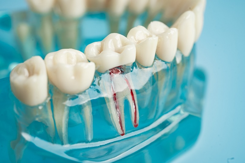
Root canal therapy carries a well-earned reputation as one of modern dentistry’s most effective treatments for saving a severely decayed or damaged tooth. For millions of patients each year, the procedure accomplishes exactly what it sets out to do: it removes acute infection, eliminates pain, and preserves the natural tooth structure for decades. Because the treatment hollows out, disinfects, and seals the inner pulp chamber—effectively removing the inflamed nerve tissue—most people assume that once a root canal is complete, that tooth is permanently immune to pain or future complications.
Yet even with advanced endodontic technology, a small percentage of root canal treatments eventually fail. A failure does not necessarily mean your dentist made an error during the original appointment. Complex root canal anatomy, microscopic accessory branches, or the gradual breakdown of an overlying dental crown can quietly invite oral bacteria back into the sealed chamber. What makes a failing root canal particularly tricky is that the interior of the tooth can no longer register sensation, yet the surrounding bone and ligament tissue remain remarkably sensitive. Learning to recognize the warning signs early can mean the difference between straightforward retreatment and losing the tooth altogether.
Why a Treated Tooth Can Still Harbor Infection
To understand why a root canal fails, it helps to look at the anatomy beneath the surface. During standard endodontic therapy, a clinician removes infected or necrotic pulp tissue, shapes and disinfects the root canals, and packs the space with an inert filling material called gutta-percha before sealing the tooth with a permanent filling or crown.
When a root canal fails, bacteria have found a way to survive inside the tooth or re-enter from the mouth. Human teeth are intricate; a single root can contain microscopic fins, lateral canals, and tiny deltas that can shelter stubborn bacteria despite rigorous irrigation. Over months or years, those remaining bacteria can slowly multiply. In other cases, a cracked filling, an ill-fitting crown margin, or delayed restoration after the initial procedure allows saliva and bacteria to seep down into the sealed canals.
Once bacteria reach the root apex—the opening at the very tip of the root—they spill directly into the surrounding periodontal ligament and jawbone. The tooth itself may lack an internal nerve, but the living bone and ligament supporting it possess an extensive network of pain receptors. This resulting chronic inflammation, known as apical periodontitis, generates the symptoms that alert patients that something is wrong.
Cardinal Signs That Your Root Canal Is Failing
Symptoms of a failing root canal do not always mirror the throbbing toothache that prompted the initial treatment. They can emerge weeks, months, or even ten years after the procedure, presenting as either subtle, intermittent nuisances or sudden, acute flare-ups.
Persistent or Late-Onset Biting Discomfort
Mild soreness for several days following a root canal is an expected part of the healing process as tissues recover from instrumentation. However, if that soreness never fully fades, or if a tooth that felt completely normal for years suddenly hurts when you chew, that sensitivity should raise a red flag.
Biting sensitivity occurs because active inflammation at the root tip creates localized pressure inside the jawbone. When you chew or tap the tooth, that mechanical force drives the root down into the inflamed pocket of tissue. If eating firm foods on that side of your mouth produces a sharp twinge or an ache that lingers after you stop chewing, the seal around the root apex is likely compromised.
The Appearance of a Fistula or Gum Pimple
One of the clearest physical indicators of an unresolved root canal infection is a small, pimple-like bump on the gum tissue near the base of the tooth. Known clinically as a parulis or fistulous tract, this bump serves as an exit channel that your body creates to vent accumulated pus from an abscess at the root tip.
Patients often notice that the bump swells, bursts, and releases a foul-tasting, slightly salty fluid, which often brings a temporary feeling of pressure relief. Because the discomfort subsides after drainage, many people mistakenly assume the infection resolved on its own. In reality, the drainage tract simply relieved hydraulic pressure while the underlying bacterial colony continues to dissolve the surrounding bone.
Localized Swelling and Tissue Tenderness
Unlike the focused drainage of a fistula, some failing root canals produce generalized swelling across the adjacent gumline. The tissue near the root may appear noticeably puffy, red, and tender to the touch, making routine brushing and flossing uncomfortable.
In more aggressive cases, swelling can extend beyond the gum tissue into the cheek, jaw, or under the chin. Any visible facial swelling is considered an urgent dental issue. It indicates that the bacterial infection is breaking through the bone and spreading into the soft tissue spaces of the face and neck, requiring prompt clinical intervention.
A Sensation That the Tooth Sits High or Feels Loose
As inflammatory fluid gathers around the root tip, it can physically push the tooth upward by a fraction of a millimeter within its socket. Consequently, you may notice that the treated tooth makes contact before any of your other teeth when you close your mouth or chew.
Over time, chronic apical inflammation causes bone resorption, gradually eating away the dense bone socket that anchors the root. If enough bone support is lost, the tooth may begin to feel unstable or slightly loose when pressed with a finger or tongue. A shifting or mobile tooth years after endodontic treatment is a strong sign of underlying bone loss.
Chronic Sinus Pressure and Drainage
Failing root canals in the upper jaw often produce symptoms that masquerade as common seasonal sinus troubles. The roots of the upper molars and premolars sit directly beneath—and in some cases extend slightly into—the floor of the maxillary sinus.
When an infection develops at the tip of these upper roots, bacteria and inflammatory byproducts can breach the thin sinus membrane. This complication, known as maxillary sinusitis of endodontic origin, typically causes unilateral congestion, sinus pressure beneath one cheekbone, postnasal drip, or an unpleasant odor in the nose that does not respond to conventional allergy treatments or decongestants.
What to Do If You Suspect a Failure
Ignoring the warning signs of a failing root canal rarely leads to an uneventful outcome. Left unaddressed, chronic infection quietly destroys jawbone structure, which not only complicates saving the natural tooth but can also jeopardize future tooth replacement options such as dental implants.
If you recognize any of these symptoms, your first step should be an evaluation by a dentist or an endodontist, a specialist focused exclusively on diagnosing and treating diseases of the dental pulp. Today’s clinicians have sophisticated tools and techniques designed to diagnose and resolve failing treatments:
-
3D Cone Beam Imaging: Traditional flat X-rays can hide tiny infections behind existing roots or dense bone. Modern endodontic offices routinely use cone beam computed tomography (CBCT) to generate high-resolution 3D views of the tooth, revealing missed canals, subtle fractures, and hidden bone lesions.
-
Non-Surgical Retreatment: In many instances, the tooth can be successfully preserved through retreatment. The endodontist carefully removes the existing restoration, cleans out the old filling material, locates previously missed canals, thoroughly disinfects the system, and places a new seal.
-
Apicoectomy: If the existing crown is intact and the canals are well sealed, but an infection persists at the very tip of the root, a minor microsurgical procedure called an apicoectomy can resolve the issue. The specialist makes a small incision in the gum, removes the infected root tip, cleans the surrounding bone cavity, and places a small filling at the end of the root.
-
Tooth Extraction and Replacement: If a tooth has sustained a non-restorable vertical root fracture or severe bone destruction, extraction may be the healthiest path forward. Once the area heals, modern solutions like dental implants or fixed bridges can restore normal function and appearance seamlessly.
Discovering renewed discomfort in a tooth you thought was permanently treated can certainly be frustrating. However, catching the issue early gives you the widest range of conservative, tooth-preserving options. Keeping an eye on subtle changes in bite comfort, gum health, and chewing sensitivity ensures you can address problems before they turn into complex dental emergencies.
